Big Picture
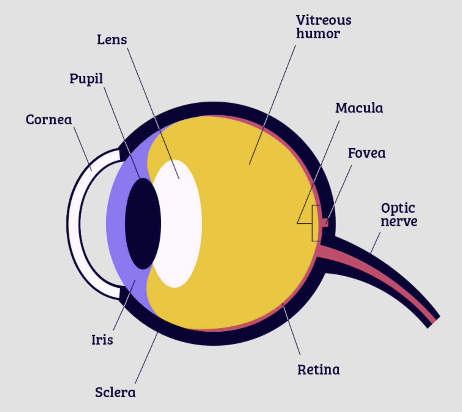
Think of your eye like a camera, but one that's alive and constantly repairing itself. Light comes in through the cornea (clear front window), gets focused by the lens, passes through the vitreous (clear gel), and lands on the retina (the sensor). The macula is the sweet spot in the center for sharp detail vision.
Quick orientation: If you're looking at someone's right eye, their nasal side is on your left. "Anterior" = front (near eyelids), "Posterior" = back (near optic nerve).
💧 Vitreous Humor, The Gel
The vitreous fills ~80% of your eye's volume. It's 99% water + 1% collagen/hyaluronic acid mesh, like a very soft Jell-O. It's crystal clear when young, firmly attached to the retina.
What happens with age: The collagen mesh shrinks (syneresis), water pockets form (liquefaction), and the gel pulls away from the retina, posterior vitreous detachment (PVD). This is normal! 75% of people over 65 have it.
Floaters = clumps of collagen casting shadows on your retina. They live in the vitreous, which is why no eye drop can reach them — the vitreous has no blood supply.
Key Facts
- No blood vessels = no immune cells = no way for drops to penetrate
- PVD is a normal aging change, not a disease
- Weiss ring = the circular floater from the optic nerve attachment
🎯 Macula, Your High-Def Zone
The macula is a tiny (~5.5 mm) yellowish spot in the center of your retina. It's packed with cones (color/detail photoreceptors) and has ZERO rods (night vision). That's why you can read this text but can't see stars in your peripheral vision.
Why it's special: Highest metabolic rate in the body. Relies on the choroid(blood vessel layer behind retina) and RPE (retinal pigment epithelium, the support crew) for nutrients and waste removal.
AMD attacks the support system: Drusen (debris) build up under the RPE → RPE struggles → photoreceptors starve → central vision fades. Wet AMD = abnormal blood vessels grow through cracks (choroidal neovascularization).
Macula vs. Peripheral Retina
| Feature | Macula | Peripheral Retina |
|---|---|---|
| Photoreceptors | Cones only (detail/color) | Rods (motion/night) |
| Ganglion cells | 1:1 (high acuity) | Many:1 (sensitivity) |
| Blood vessels | None in center (fovea) | Rich supply |
| Damage effect | Central blur/blind spot | Side vision loss |
🛢️ Meibomian Glands, The Oil Factories
You have ~25–40 of these in your upper lid and ~20–30 in the lower lid. They run vertically along the lid margin, opening right at the lash line.
Job: Pump out meibum (oil) every time you blink. This forms the lipid layerof your tear film, the top coat that stops tears from evaporating in seconds.
MGD (Meibomian Gland Dysfunction): Oil thickens (like toothpaste instead of olive oil), glands clog, then atrophy. Result: tears evaporate → dry, burning, blurry eyes. This is 86% of dry eye.
Why warm compresses work: Heat melts the waxy meibum back to liquid so it can flow. That's why 10 minutes 2×/day is the gold standard — it's physics, not magic.
MGD = The Root of Most "Dry Eye"
- Glands don't regenerate once atrophied — early treatment saves them
- Lipid tears (Refresh Mega-3, Systane Complete) replace what glands can't
- Omega-3 rTG improves oil quality from the inside (6–12 weeks)
- IPL/LipiFlow = physical unclogging for stubborn cases
🔍 Lens, The Flexible Focus
The lens sits behind the iris, suspended by zonules (tiny fibers) attached to the ciliary body. It's made of layered crystallin proteins, transparent, flexible, and continually growing throughout life.
Accommodation: Ciliary muscle contracts → zonules relax → lens gets rounder → near focus. Young lens = soft & flexible. Age 40+ = proteins cross-link, stiffen → presbyopia.
Cataract: Those same proteins eventually clump and yellow. The lens loses transparency. No drop reverses this — the proteins are covalently cross-linked. Surgery replaces the lens with an IOL.
Presbyopia ≠ "Weak Eyes"
- It's universal — starts ~40, done ~55
- NOT caused by "not exercising your eyes"
- Reading glasses / Vuity drops / premium IOLs = optical workarounds
🎨 Retina, RPE & Choroid, The Support Team
Retina (Neural Layer)
Photoreceptors (rods/cones) → bipolar cells → ganglion cells → optic nerve. Brain tissue, basically.
RPE (Retinal Pigment Epithelium)
Single layer of support cells. Feeds photoreceptors, recycles visual pigment, hauls away trash. First to fail in AMD.
Choroid (Blood Supply)
High-flow vascular bed behind RPE. Delivers oxygen/nutrients, removes heat. Fastest blood flow per gram in body.
The Cheat Sheet
Vitreous — Floaters live here. No drops reach it.
Macula — Central vision HQ. AREDS2 helps intermediate AMD.
Meibomian glands — Oil factories in the lids. MGD = most dry eye.
Lens — Focuses light. Stiffens with age. Clouds → cataract surgery.
Retina / RPE / Choroid — Sensor + support + fuel line. RPE fails first in AMD.
Cornea — Clear front window. Needs tears to stay smooth.